The Kidneys
Written by Oliver Jones
Last updated April 16, 2024 • 62 Revisions •
The kidneys are bilateral bean-shaped organs, reddish-brown in colour and located in the posterior abdomen. Their main function is to filter and excrete waste products from the blood. They are also responsible for water and electrolyte balance in the body.
Metabolic waste and excess electrolytes are excreted by the kidneys to form urine . Urine is transported from the kidneys to the bladder by the ureters . It leaves the body via the urethra , which opens out into the perineum in the female and passes through the penis in the male.
In this article we shall look at the anatomy of the kidneys – their anatomical position, internal structure and vasculature.
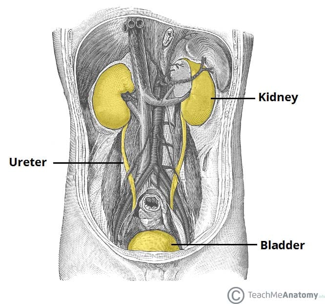
Fig 1 Overview of the urinary tract.

Premium Feature
Pro feature, anatomical position.
The kidneys lie retroperitoneally (behind the peritoneum) in the abdomen, either side of the vertebral column.
They typically extend from T12 to L3 , although the right kidney is often situated slightly lower due to the presence of the liver. Each kidney is approximately three vertebrae in length.
The adrenal glands sit immediately superior to the kidneys within a separate envelope of the renal fascia .
Dissection Images

Kidney Structure
The kidneys are encased in complex layers of fascia and fat. They are arranged as follows (deep to superficial):
- Renal capsule – tough fibrous capsule.
- Perirenal fat – collection of extraperitoneal fat.
- Renal fascia (also known as Gerota’s fascia or perirenal fascia) – encloses the kidneys and the suprarenal glands.
- Pararenal fat – mainly located on the posterolateral aspect of the kidney.
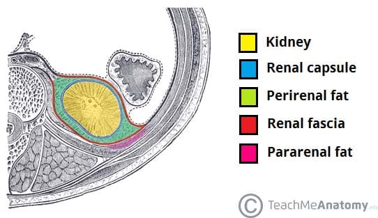
Fig 2 The external coverings of the kidney.
Internally, the kidneys have an intricate and unique structure. The renal parenchyma can be divided into two main areas – the outer cortex and inner medulla . The cortex extends into the medulla, dividing it into triangular shapes – these are known as renal pyramids .
The apex of a renal pyramid is called a renal papilla . Each renal papilla is associated with a structure known as the minor calyx , which collects urine from the pyramids. Several minor calices merge to form a major calyx . Urine passes through the major calices into the renal pelvis , a flattened and funnel-shaped structure. From the renal pelvis, urine drains into the ureter, which transports it to the bladder for storage.
The medial margin of each kidney is marked by a deep fissure, known as the renal hilum . This acts as a gateway to the kidney – normally the renal vessels and ureter enter/exit the kidney via this structure.
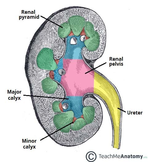
Fig 3 The internal structure of the kidney.
Anatomical Relations
The kidneys sit in close proximity to many other abdominal structures which are important to be aware of clinically:
Arterial Supply
The kidneys are supplied with blood via the renal arteries , which arise directly from the abdominal aorta, immediately distal to the origin of the superior mesenteric artery . Due to the anatomical position of the abdominal aorta (slightly to the left of the midline), the right renal artery is longer, and crosses the vena cava posteriorly.
The renal artery enters the kidney via the renal hilum. At the hilum level, the renal artery forms an anterior and a posterior division, which carry 75% and 25% of the blood supply to the kidney, respectively. Five segmental arteries originate from these two divisions.
The avascular plane of the kidney (line of Brodel) is an imaginary line along the lateral and slightly posterior border of the kidney, which delineates the segments of the kidney supplied by the anterior and posterior divisions. It is an important access route for both open and endoscopic surgical access of the kidney, as it minimises the risk of damage to major arterial branches.
Note: The renal artery branches are anatomical end arteries – there is no communication between vessels. This is of crucial importance; as trauma or obstruction in one arterial branch will eventually lead to ischaemia and necrosis of the renal parenchyma supplied by this vessel.
The segmental branches of the renal undergo further divisions to supply the renal parenchyma:
- Each segmental artery divides to form interlobar arteries . They are situated either side every renal pyramid.
- These interlobar arteries undergo further division to form the arcuate arteries .
- At 90 degrees to the arcuate arteries, the interlobular arteries arise.
- The interlobular arteries pass through the cortex, dividing one last time to form afferent arteriole s .
- The afferent arterioles form a capillary network, the glomerulus, where filtration takes place. The capillaries come together to form the efferent arterioles.
In the outer two-thirds of the renal cortex, the efferent arterioles form what is a known as a peritubular network , supplying the nephron tubules with oxygen and nutrients. The inner third of the cortex and the medulla are supplied by long, straight arteries called vasa recta.
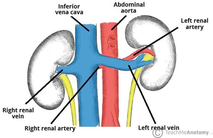
Fig 4 Arterial and venous supply to the kidneys.
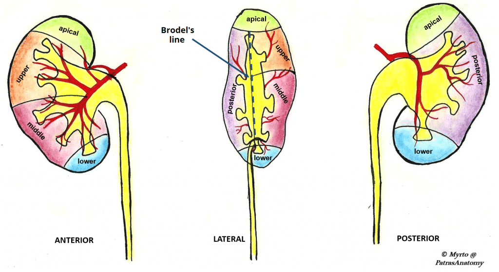
Fig 5 Arterial supply to the kidney can be divided into five segments.
Clinical Relevance
Variation in arterial supply to the kidney.
The kidneys present a great variety in arterial supply; these variations may be explained by the ascending course of the kidney in the retroperitoneal space , from the original embryological site of formation (pelvis) to the final destination (lumbar area). During this course, the kidneys are supplied by consecutive branches of the iliac vessels and the aorta.
Usually the lower branches become atrophic and vanish while new, higher ones supply the kidney during its ascent. Accessory arteries are common (in about 25% of patients). An accessory artery is any supernumerary artery that reaches the kidney. If a supernumerary artery does not enter the kidney through the hilum, it is called aberrant .
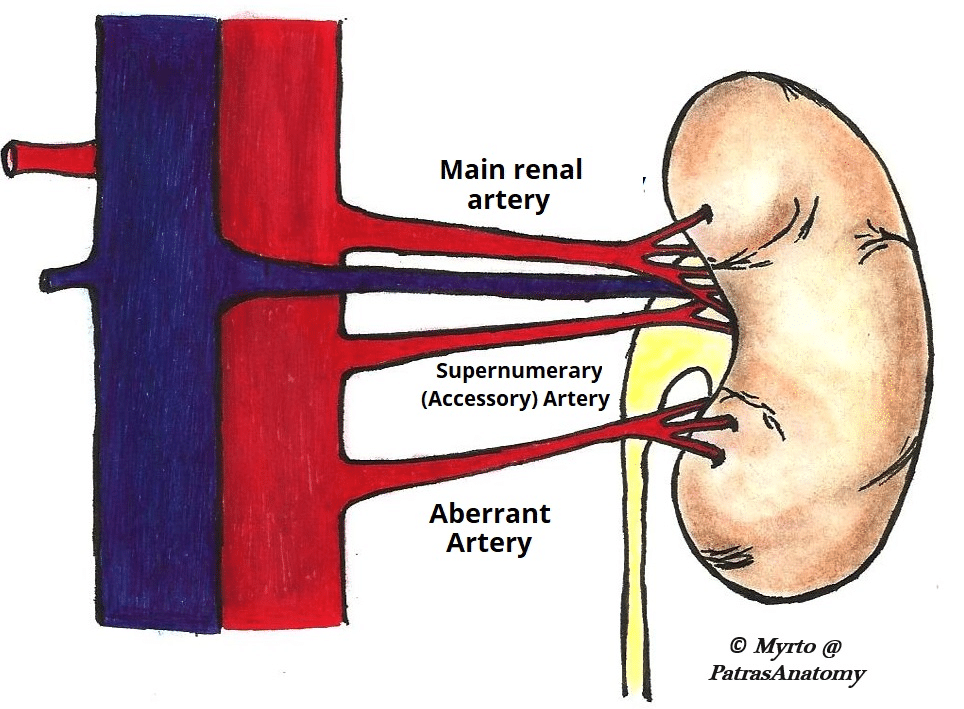
Fig 6- Supernumerary arteries of the kidney,
Venous Drainage
The kidneys are drained of venous blood by the left and right renal veins . They leave the renal hilum anteriorly to the renal arteries, and empty directly into the inferior vena cava.
As the vena cava lies slightly to the right, the left renal vein is longer, and travels anteriorly to the abdominal aorta below the origin of the superior mesenteric artery. The right renal artery lies posterior to the inferior vena cava.
Lymph from the kidney drains into the lateral aortic (or para-aortic) lymph nodes , which are located at the origin of the renal arteries.
Congenital Abnormalities of the Kidneys
Pelvic kidney.
In utero, the kidneys develop in the pelvic region and ascend to the lumbar retroperitoneal area. Occasionally, one of the kidneys can fail to ascend and remains in the pelvis – usually at the level of the common iliac artery.
Horseshoe Kidney
A horseshoe kidney (also known as a cake kidney or fused kidney) is where the two developing kidneys fuse into a single horseshoe-shaped structure.
This occurs if the kidneys become too close together during their ascent and rotation from the pelvis to the abdomen – they become fused at their lower poles (the isthmus ) and consequently become ‘stuck’ underneath the inferior mesenteric artery .
This type of kidney is still drained by two ureters (although the pelvices and ureters remain anteriorly due to incomplete rotation) and is usually asymptomatic, although it can be prone to obstruction .
Renal Cell Carcinoma
The kidney is often the site of tumor development, most commonly renal cell carcinoma.
Due to the segmental vascular supply of the kidney it is often feasible to ligate the relative arteries and veins and remove the tumour with a safe zone of healthy surrounding parenchyma ( partial nephrectomy ) without removing the entire kidney or compromising its total vascular supply by ischaemia.
Do you think you’re ready? Take the quiz below
Question 1 of 3

Recommended Reading
Forgot password.
Please enter your username or email address below. You will receive a link to create a new password via emai and please check that the email hasn't been delivered into your spam folder.
We use cookies to improve your experience on our site and to show you relevant advertising. To find out more, read our privacy policy .
Privacy Overview
Report question, rate this article.
Kidney: Structure, Function and Related Diseases
The structure of human kidney can be seen as two reddish bean-shaped organs that are located below the rib cage on each side of the spine. They are almost a fistful in size, measuring around 10-12cm. Kidneys are the main organs in the human excretory system, which takes part in the filtration of the blood before the urine is formed. Let us look at the structure of the organ for a better understanding.
External and Internal Features of Kidney
- It has a convex and concave border.
- Towards the inner concave side, a notch called the hilum is present through which the renal artery enters the kidney and the renal vein and ureter leave.
- The outer layer of the kidney is a tough capsule.
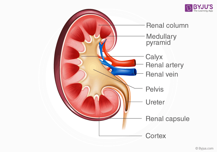
- On the inside, the kidney is divided into an outer renal cortex and an inner renal medulla.
- The hilum extends inside the kidney into a funnel-like space called the renal pelvis.
- The renal pelvis has projections called calyces(sing: calyx).
- The medulla is divided into medullary pyramids, which project into the calyces.
- Between the medullary pyramids, the cortex extends as renal columns called Columns of Bertini.
- The kidney is made up of millions of smaller units called nephrons which are also the functional units.
Read: Nephrons
A simple structure of kidney can be understood by the following diagram:
- The most important function of the kidney is to filter the blood for urine formation.
- It excretes metabolic wastes like urea and uric into the urine.
- Erythropoietin: It is released in response to hypoxia
- Renin: It controls blood pressure by regulation of angiotensin and aldosterone
- Calcitriol: It helps in the absorption of calcium in the intestines
- It maintains the acid-base balance of the body by reabsorbing bicarbonate from urine and excreting hydrogen ions and acid ions into the urine.
- It also maintains the water and salt levels of the body by working together with the pituitary gland .
Read: Urine Formation
Diseases related to Kidneys
In uremia, the kidneys are damaged, and there is a buildup of urea and other toxins in the blood, which is fatal and can cause kidney failure. Patients may experience fatigue, itching, muscle twitching, and loss of mental concentration. The urea can be removed by the process of hemodialysis.
2. Renal Calculi
Commonly called kidney stones, these are deposits of salt and minerals in our body. Symptoms include severe abdominal pain and nausea. The stones can be dissolved with medicines, or it passes with urine by improving diet and water intake.
3. Glomerulonephritis
It is the inflammation of the glomerulus. Symptoms include pink urine, oedema or swelling on the face and high blood pressure. It requires medical attention for prevention.
Note: Any person with high blood pressure and diabetes is more prone to kidney related diseases.
Explore BYJU’S Biology to learn more.
- Facts about Kidneys
- Regulation Of Kidney Function
- Kidney Function Test
- Kidney Failure Symptoms
- Difference Between Left and Right Kidney
What are the first signs of kidney problems?
You can see blood in your urine and it is also foamy. You will have a problem concentrating on things and will face swelling and dryness on your skin.
Can you live without a kidney?
It is not possible to live without a kidney. But since we have two of them, it is possible to survive with one kidney.
What causes kidney problems?
People with diseases like diabetes and high blood pressure have a high chance of developing kidney problems.
Leave a Comment Cancel reply
Your Mobile number and Email id will not be published. Required fields are marked *
Request OTP on Voice Call
Post My Comment
Thanking you Team Biju’s for this knowledge free of cost 😊
Register with BYJU'S & Download Free PDFs
Register with byju's & watch live videos.

IMAGES
VIDEO
COMMENTS
The kidneys are bilateral organs placed retroperitoneally in the upper left and right abdominal quadrants and are part of the urinary system. Their shape resembles a bean, where we can describe the superior and inferior …
The kidneys are two bilateral bean shaped organs, located in the posterior abdomen. They are reddish-brown in colour. In this article we shall look at the anatomy of the kidneys - their anatomical position, internal structure …
Each kidney is surrounded by a renal capsule, an adipose capsule and renal fascia. Production of renin activates the renin-angiotensin-aldosterone system in the regulation of blood pressure. …
The kidney has a wide array of responsibilities that range from regulation of fluid volume and osmolarity, management of electrolytes, …
This Osmosis High-Yield Note provides an overview of Anatomy and Physiology of the Renal System essentials. All Osmosis Notes are clearly laid-out and contain striking images, tables, and diagrams to help visual learners …
At the medial margin of each kidney lies the renal hilum, where the renal artery enters, and the renal pelvis and vein leave the renal sinus. The renal vein is found anterior to the renal artery, which is anterior to the renal pelvis. …
Overview. The kidneys are two bean-shaped organs responsible for filtering minerals from the blood, maintaining overall fluid balance, excreting waste products, and regulating blood volume to...
Assessing renal function is crucial in treating patients with kidney disease or pathologies affecting renal function. Renal function tests are useful for identifying the presence of renal disease, monitoring the response of …
Kidneys are the main organs in the human excretory system, which takes part in the filtration of the blood before the urine is formed. Let us look at the structure of the organ for a better …